
Skin Cancer vs Dark Spots: How to Tell the Difference and When to Worry
Skin Cancer vs Dark Spots: How to Tell the Difference and When to Worry
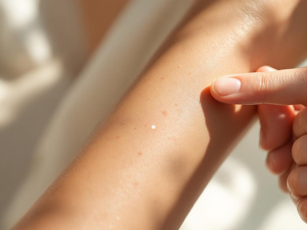
You spotted something on your forearm this morning. A brown mark you don't remember seeing before — or maybe one you've had for years that suddenly looks different in the bathroom light. Within thirty seconds, you've typed skin cancer vs dark spots into your phone and fallen into the rabbit hole every dermatologist's patient describes the same way: equal parts panic and paralysis. That anxiety is rational. Melanoma is the most lethal form of skin cancer. But here is the part the search results rarely lead with — the overwhelming majority of pigmented marks on adult skin are completely benign.
The difference between a spot that needs a same-week dermatology visit and one that can be monitored at home comes down to a single distinction. Dark spots are about pigment. Skin cancer is about cellular behavior. A solar lentigo — what most people call an age spot — is melanin clustered in the upper layers of skin from years of sun exposure. Melanoma is the pigment-producing cells themselves replicating without regulation. One is a cosmetic concern. The other is a medical emergency. The visual signs that separate them are learnable in about ten minutes of focused reading, which is why this article exists.
The stakes justify the time. According to the American Cancer Society's Cancer Facts & Figures 2025, roughly 104,960 new invasive melanoma cases are projected in the United States this year alone. The National Cancer Institute's SEER program reports that 5-year relative survival for localized melanoma sits near 99%, but drops to roughly 35% for distant-stage disease. That gap — the difference between a minor outpatient procedure and a life-altering oncology pathway — is largely determined by how early the lesion is identified. Visual literacy about your own skin is, statistically, one of the highest-leverage health habits you can build.
By the end of this read, you will be able to apply the ABCDE rule the way a dermatologist's intake nurse does, recognize the most common benign mimics by name and visual signature, match your specific situation to the correct professional and the correct timeline, and run a monthly self-check that catches change at the stage where treatment is still simple. None of this is diagnosis. Anyone with genuine doubt should see a board-certified dermatologist. What follows is structured education — a framework that converts vague worry into specific decisions.
Dark spots are about pigment sitting in the skin. Skin cancer is about cells that have stopped following the rules.
Table of Contents
- Why Dark Spots Trigger the Cancer Question
- The ABCDE Rule: Five Visual Signals That Earn a Closer Look
- The Benign Look-Alikes: Pigmentation That Isn't Cancer
- How Melanoma Behaves Over Time
- Who to Call and How Fast
- The Monthly Skin Self-Check
- Real Scenarios, Real Decisions
The ABCDE Rule: Five Visual Signals That Earn a Closer Look
The ABCDE rule was developed in 1985 by dermatologists Robert Friedman, Darrell Rigel, and Alfred Kopf at NYU School of Medicine, originally as ABCD and later expanded to include "E" for Evolving. It is the most widely adopted public-facing melanoma screening framework, endorsed by both the American Academy of Dermatology and the Skin Cancer Foundation. Its purpose is narrow and useful: help non-experts decide whether a pigmented mark deserves professional eyes.

A — Asymmetry. Draw an imaginary line through the middle of the spot. If the two halves do not mirror each other, that is asymmetry. Benign moles and age spots grow uniformly, so they tend to be symmetrical. Melanoma grows in unpredictable directions because the cells are not following normal regulatory signals.
B — Border irregularity. Benign spots have smooth, well-defined edges. Melanoma borders are often notched, scalloped, blurred, or fade unevenly into surrounding skin. The irregularity reflects aggressive, uneven cellular invasion at the lesion's edge.
C — Color variation. Benign age spots are typically a single shade of brown or tan throughout. Melanoma frequently shows multiple colors within one lesion — tan, dark brown, black, sometimes red, white, or blue-gray. This reflects melanin being produced inconsistently and at different skin depths, with possible areas of immune-driven regression.
D — Diameter greater than 6mm. That is roughly the width of a standard pencil eraser. Treat this as a threshold of suspicion, not a rule. Melanomas can be smaller, and benign spots can be larger. Size combined with any other ABCDE flag is what raises the urgency level.
E — Evolving. Any change in size, shape, color, elevation, or the appearance of new symptoms — itching, bleeding, crusting — over a span of weeks to months. The AAD considers E the single most important warning sign because benign spots are almost always stable over time.
The ABCDE rule is a triage tool, not a diagnostic test. A spot that fails one criterion may still be benign. A spot that fails three or more should be examined by a dermatologist within days, not weeks. The rule also has known limitations — it can miss nodular melanomas, which are often symmetrical and uniformly colored, and amelanotic melanomas, which lack pigment entirely. Treat ABCDE as a starting point for understanding skin cancer warning signs, not as a clearance. When the question is how to tell if a mole is cancerous, ABCDE answers half of it. The other half is behavior over time, which the next sections cover.
The Benign Look-Alikes: Pigmentation That Isn't Cancer
Most pigmented marks people worry about are one of five common benign types. Knowing them by name and visual signature reduces anxiety and — equally important — sharpens your ability to identify when something does not fit any benign pattern.
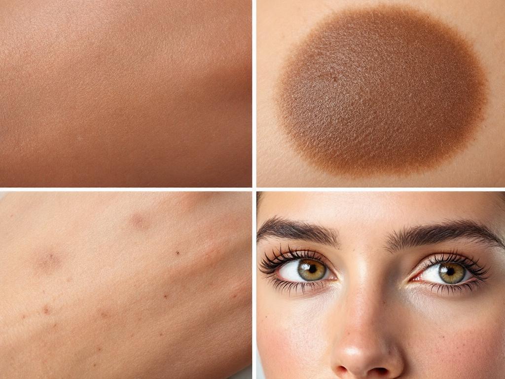
Solar Lentigines (Age Spots / Sun Spots). Flat, uniform tan to dark brown, sharply demarcated edges, typically 2–20mm in diameter. Most common on hands, face, shoulders, and upper back — the cumulative-sun-exposure zones. According to the American Academy of Dermatology, they typically appear from age 40 onward and are caused by UV-driven melanin clustering in the upper skin. They carry zero malignancy risk and remain stable for years; the age spots and hyperpigmentation treatment pathway is purely cosmetic. The red flag is not the spot itself — it is when a spot you have had for a decade suddenly starts changing.
Melasma. Larger, blotchy, symmetrical patches typically distributed across the cheeks, forehead, upper lip, or jawline. Driven by hormones (pregnancy, oral contraceptives, hormonal therapy) combined with UV exposure, predominantly affecting women aged 20–50. The biological pathway is entirely distinct from melanoma — melasma involves overactive melanocytes producing excess pigment in response to hormonal and UV signals, not malignant transformation. Treatment combines topical agents (azelaic acid, tranexamic acid, hydroquinone under guidance), professional peels, and rigorous sun protection.
Post-Inflammatory Hyperpigmentation (PIH). Brown, gray-brown, or pink-brown marks that appear precisely where acne, eczema, ingrown hairs, or any skin injury has occurred. Most common in skin of color. PIH typically resolves over 3–24 months with sun protection and a post-acne discoloration treatment protocol. The defining feature, and the one that should anchor your mental model: PIH fades over time. Melanoma does not fade. If a mark you have been treating for post-acne discoloration is darkening rather than lightening month after month, that pattern needs professional evaluation.
Seborrheic Keratosis. Waxy, slightly raised, "stuck-on" appearance — often described as looking like a piece of brown candle wax pressed onto the skin surface. Color ranges from light tan to nearly black. Common in adults over 50. Per the AAD, they are entirely benign overgrowths of immature skin cells. They are frequently mistaken for melanoma because they can appear dark and irregularly shaped. The waxy, raised, "could almost be peeled off" texture is the giveaway that distinguishes them from a flat pigmented lesion of concern. Often this rough, raised surface overlaps with broader skin texture concerns that clients want addressed alongside pigmentation.
Freckles (Ephélides). Small (1–2mm), flat, uniformly tan or light brown, scattered in clusters on sun-exposed areas. Genetic in origin — driven by MC1R gene variants more common in fair-skinned individuals. They darken in summer and lighten in winter. That seasonal cycle alone distinguishes them from any concerning pigment change, since melanoma does not respond to seasonal UV the way freckles do.
A spot that fits cleanly into one of these five categories — and has been stable — is almost never cancer. A spot that fits none of them, or that fits but has started behaving differently, deserves professional eyes. That distinction between melanoma vs age spot is, in clinical practice, more about behavior than appearance, which is why the next section matters.
How Melanoma Behaves Over Time
The ABCDE rule captures a snapshot. But melanoma's most reliable signal is behavior over time. A static photograph of a lesion tells you less than a series of photographs taken across months. This section covers what those changes look like and why they matter biologically.
The evolution criterion in depth. The "E" was added to the original ABCD framework precisely because clinicians realized that change is the strongest single predictor of malignancy. Per the Skin Cancer Foundation, evolution can include darkening, lightening, expanding, becoming raised, developing satellite spots nearby, or new symptoms such as itching, tenderness, or bleeding. Timeline matters: a noticeable change over 4–8 weeks is more concerning than a slow shift over years, although slow-growing lentigo maligna melanoma is the clinical exception that proves the rule and is itself a recognized subtype.
Color complexity as a cellular signature. Benign spots are typically one or two shades because melanin is being deposited uniformly. Melanoma frequently shows three or more colors — tan, brown, black, with possible red, white, or blue-gray patches — because the cancer involves disordered melanin production at varying skin depths and areas of regression where the immune system has partially attacked the lesion. Multi-color appearance in a single spot is one of the strongest visual predictors a non-expert can recognize.
Bleeding, crusting, itching. A spot that bleeds without trauma, oozes, develops a persistent crust, or itches without an obvious cause is signaling surface disruption. These symptoms are not always present in early melanoma — many early melanomas are asymptomatic — but their appearance in any pigmented lesion warrants prompt evaluation. Per AAD guidance, persistent bleeding from a mole is a same-week dermatology visit, not a wait-and-see scenario.
Size as a risk escalator, not a rule. The 6mm threshold is a guide. Melanomas under 6mm exist, especially in younger patients or with certain subtypes, and benign moles over 6mm are common. The clinically meaningful pattern is size combined with any other ABCDE flag. A 4mm spot that is asymmetrical and changing is more concerning than a 10mm spot that has been stable and uniform for a decade.
A spot that hasn't changed in five years is almost never melanoma. A spot that changed noticeably in five weeks deserves a dermatologist within days, not months.
The ugly duckling sign. Most people's moles share a family resemblance — similar size, color, and shape. The ugly duckling is the mole that does not match the rest. The AAD endorses this as a complementary screening principle alongside ABCDE because the human eye is excellent at pattern recognition once it knows what to look for. The outlier deserves attention even if it does not independently fail any ABCDE criterion. In practice, the ugly duckling sign catches some early melanomas that ABCDE alone misses, and it requires no measurement — only honest comparison.
High-risk anatomical locations. Acral lentiginous melanoma — melanoma occurring on palms, soles, under fingernails, or under toenails — is less common overall but has worse outcomes due to delayed diagnosis. It is also disproportionately common in people with darker skin tones, where it is often missed because melanoma awareness messaging has historically focused on fair-skinned populations. A new dark streak under a nail, especially one that widens over time, a dark spot on a palm or sole, or any non-healing dark mark in these locations warrants prompt evaluation regardless of how small it is.
The diagnostic disparity. Per the Skin Cancer Foundation, 5-year melanoma survival rates are significantly lower in Black patients (around 71%) compared to white patients (around 94%), largely driven by later-stage diagnosis. The takeaway for readers of every skin tone: melanoma is less common in darker skin, but when it does occur, it is more often dangerous because it is caught later. The ABCDE rule applies regardless of skin tone, and high-risk-location vigilance — palms, soles, nail beds, mucous membranes — is especially important. If you have darker skin and have been told melanoma "isn't really a concern" for you, that messaging is statistically outdated and clinically incorrect.
Who to Call and How Fast
Different scenarios call for different professionals on different timelines. The matrix below maps the most common situations to the appropriate response and helps clarify when to see a dermatologist for dark spots versus when a cosmetology consultation is the better starting point.
| Scenario | Urgency | First Step | Why |
|---|---|---|---|
| 3+ ABCDE flags AND change in past 4 weeks | Same week | Board-certified dermatologist (in-person) | Possible melanoma; needs clinical exam and likely biopsy |
| Spot bleeding, oozing, or persistently itching | Within 48 hours | Dermatologist (in-person) | Surface disruption is high-priority warning |
| New dark streak under nail, on palm, or sole | Within 1 week | Dermatologist (in-person) | High-risk location for acral melanoma |
| 1–2 ABCDE flags, stable for 6+ months | Within 2–4 weeks | Dermatologist (in-person or telehealth) | Low immediate risk; professional confirmation needed |
| Multiple dark spots, no individual flags | Within 1–3 months | Dermatologist + cosmetologist | Baseline mole-mapping plus cosmetic strategy |
| Confirmed-benign pigmentation, cosmetic concern | Elective | Cosmetologist (online or in-person) | No medical concern; treatment optimization |
| Uncertain mark, no clear flags, want a screen | Within 2 weeks | Photo-based consult OR dermatologist | Initial triage to separate benign from suspicious |
Why a dermatologist for suspicious cases. Board-certified dermatologists are the only professionals trained to clinically evaluate, perform dermoscopy on, and biopsy suspicious lesions. No cosmetologist, aesthetician, or telemedicine service can diagnose skin cancer. If a lesion is suspicious, the path is dermatologist → biopsy → pathology. Anything else introduces delay, and delay is the variable most directly tied to outcome.
Why a cosmetologist for confirmed-benign cases. Once cancer has been ruled out, treatment becomes a cosmetic and dermatological optimization question — and remote, photo-based cosmetology consultations can be highly effective for confirmed-benign pigmentation. They are particularly well-suited for post-acne discoloration, age spots, and melasma management, where the work involves sustained protocols — topical agents, sun protection, lifestyle adjustments — rather than acute intervention. Photo-based consultations are also effective for ongoing follow-up for chronic concerns like rosacea, PIH, and early aging signs, where bi-weekly check-ins outperform episodic visits.
The role of remote-first models. Photo-based consultations work well for three things: confirmed-benign pigmentation treatment planning, initial screening to identify whether a spot's features warrant in-person dermatology referral, and ongoing follow-up for chronic concerns. They are not replacements for in-person dermoscopy or biopsy when there is suspicion of malignancy. Used in the right context, they reduce friction, improve protocol adherence, and free in-person dermatology slots for the cases that genuinely need them.
The default rule. When in doubt, see a dermatologist. A dermatology consult is rarely wasted — at minimum it establishes a baseline, and at best it catches something early that self-assessment missed. The cost of an unnecessary dermatology visit is small. The cost of a delayed melanoma diagnosis can be measured in five-year survival percentages.
The Monthly Skin Self-Check
Per the AAD, monthly self-skin-exams combined with annual dermatologist visits for higher-risk individuals are the most evidence-supported early-detection strategy a person can practice at home. The goal is not to diagnose. It is to know your baseline so well that any change becomes obvious.
The five-step monthly process.
Pick a fixed day. First Sunday of the month, the first of the month, the day you pay rent — anything you will remember without prompting. Consistency beats sophistication every time.
Set up consistent conditions. Same room, same light source, same time of day. Natural daylight near a window is ideal because it reveals color variation more accurately than warm indoor lighting. A full-length mirror plus a hand mirror covers most of the body. A partner or a smartphone on a small tripod handles the back, scalp, and posterior thighs.
Check head to toe systematically. Scalp (use a comb to part hair in sections), face, neck, ears, chest, abdomen, arms including underarms and palms, back, buttocks, legs front and back, feet including soles and between toes, nail beds. Do not skip areas the sun rarely sees. Melanoma can occur anywhere there are melanocytes, including mucous membranes and the soles of the feet.
Document with photos. Use a smartphone in consistent lighting. Capture wide regional shots and close-ups of individual spots. Place a coin or ruler beside larger spots for size reference. Save in a dated folder labeled by month. This archive is the single most useful thing you can hand a dermatologist if a question ever arises.
Compare to prior months. Look at this month's photos beside last month's. Note new spots, size changes, color changes, border changes, and any new symptoms. The comparison is where the value is — month-by-month photos turn vague impressions into objective evidence.
Escalation checklist — when to stop monitoring and make the call.
- A spot changed noticeably in less than 4 weeks → dermatologist within days
- A spot now shows 3+ ABCDE features → dermatologist within 1 week
- A spot is bleeding, oozing, or persistently itchy → dermatologist within 48 hours
- A new dark mark appeared on a nail bed, palm, or sole → dermatologist within 1 week
- A mole looks different from all your other moles ("ugly duckling") → dermatologist within 2 weeks
- Multiple new spots appeared in a short window → dermatologist for full skin screening
- You are uncertain whether a change is real → photograph it now, recheck in 2 weeks; if still uncertain, book a consult
You see your skin every day. A dermatologist sees it once or twice a year. Your baseline awareness is the most underrated early-warning tool in skin cancer detection.
Why this beats episodic checking. Per the Skin Cancer Foundation, people who self-examine monthly detect melanomas at earlier, more treatable stages. The mechanism is baseline familiarity. You cannot notice change without knowing what was there before, and a once-a-year glance in the mirror is not enough scaffolding to catch a lesion that evolved over six weeks. Monthly self-checks pair well with early signs of aging prevention protocols because both depend on the same underlying habit — consistent, structured attention to your skin over time rather than reactive panic when something catches your eye.
Real Scenarios, Real Decisions
The framework is more memorable when applied to specific situations. Below are seven common presentations readers may recognize, with the most likely diagnosis and the immediate action.
| Your Situation | Most Likely | Immediate Action | Reasoning |
|---|---|---|---|
| Flat brown spot on hand, 5+ years, uniform, no change | Solar lentigo | Cosmetic treatment optional | Stability + uniformity rules out melanoma |
| Brown mark after a pimple cleared, slowly fading | Post-inflammatory hyperpigmentation | Sun protection + topical plan | Fading is the hallmark; melanoma does not fade |
| Symmetric blotchy patches on cheeks, hormonal trigger | Melasma | Cosmetic treatment + sun protection | Hormonal pattern + symmetric distribution |
| Asymmetrical patch on cheek, darkened in 6–8 weeks, itchy | Possible melanoma | Dermatologist within 1 week | Itching + rapid darkening is a red-flag combination |
| New small mole on back, symmetric, single color, stable 3 months | Likely benign acquired mole | Photograph and monitor monthly | New moles can appear into the 30s |
| Irregular spot, multiple colors, expanded in 6 weeks, crusty | High suspicion for melanoma | Dermatologist this week | Multiple ABCDE flags + rapid evolution |
| Dark streak under fingernail or toenail, widening | Possible acral melanoma | Dermatologist within 1 week | High-risk location for aggressive subtype |
The most common pattern in this table — and in real clinical life — is that change over time plus visual complexity is the differentiator. Static, uniform, predictable spots are almost always benign, regardless of how dark they appear. Spots that are evolving, multi-colored, or symptomatic deserve professional attention even when they are small. That single mental model — stable and uniform versus changing and complex — does most of the work of separating skin cancer vs dark spots in everyday situations.
A few practical guidelines emerge from these scenarios.
When the situation is clearly benign — a long-stable spot in a classic sun-exposure location with uniform appearance — the decision is purely cosmetic. Treatment options range from topical brighteners (azelaic acid, tranexamic acid, vitamin C, hydroquinone under professional guidance) to chemical peels and targeted laser. A remote cosmetology consultation can build a personalized protocol around your skin type, history, and tolerance, which matters because the same active ingredient that works on resilient skin can flare a sensitive complexion. Clients with reactive baselines often need a parallel sensitive skin care approach layered into any pigmentation protocol.
When the situation involves any uncertainty — the spot does not fit a clean benign pattern, or it is behaving in ways you cannot explain — the answer is always a dermatologist first, cosmetologist second. The cost of an unnecessary dermatology visit is small. The cost of a delayed melanoma diagnosis is enormous. This is the situation where knowing when to see a dermatologist for dark spots matters most: the answer is now, not after another month of monitoring.
When the situation involves clear red flags — rapid change, multiple colors, bleeding, high-risk location — speed matters. Same-week dermatology contact is the standard. Per NCI SEER data, the survival gap between localized and distant-stage melanoma is one of the steepest in oncology, and the variable most under your control is how quickly you act when something looks wrong. Time saved at the front end translates directly to outcome at the back end.
For everything that lands in the confirmed-benign category — the post-acne marks, the age spots, the melasma patches, the rosacea-related discoloration — that is where consistent skincare protocols, professional-grade topicals, and ongoing follow-up genuinely change outcomes. Acne treatment protocols, for instance, work best when paired with a parallel plan for the discoloration acne leaves behind, because treating one without the other tends to leave clients frustrated even when the active breakouts have cleared. That is the work of cosmetology, and remote-first models make it accessible without the friction of repeated in-person appointments — which, for chronic and slow-resolving concerns, is often the difference between a protocol completed and a protocol abandoned.